Oakstone UCSF Neuro and Musculoskeletal Imaging 2026
Oakstone UCSF Neuro and Musculoskeletal Imaging 2026
Regular price
$35.00 USD
Regular price
$35.00 USD
Sale price
Unit price
per
Couldn't load pickup availability

Oakstone UCSF Neuro and Musculoskeletal Imaging 2026
Regular price
$35.00
Regular price
$35.00
Sale price
Unit price
per
Oakstone UCSF Neuro and Musculoskeletal Imaging 2026
+ Target Audience: radiologists + Information:State-of-the-Art Radiology Insights
UCSF Neuro and Musculoskeletal Imaging is a comprehensive, online video update of clinically relevant topics in neuroradiology and musculoskeletal imaging, including a discussion of optimized protocols and review of indications commonly seen in general practice. Continuing medical education lectures focus on boosting diagnostic confidence and improving communication when referring cases to clinicians and specialists. Key topics include:- Neuroradiologic emergencies in adult and pediatric patients
- Head and neck masses, mimics, and post-treatment changes
- Headache evaluation, cranial nerve imaging, and skull base lesions
- Neonatal brain development, injury, and autoimmune disease
- Spine imaging, inflammatory lesions, tumors, and reporting strategies
- Musculoskeletal joint imaging, injuries, and internal derangements
- Arthritis, metabolic bone disease, and infection imaging
- Musculoskeletal ultrasound applications as a problem-solving tool
Learning Objectives
At the conclusion of this activity, the participant will be able to:- Recommend CT/MR protocols for brain, head & neck, spine, nerve, and musculoskeletal imaging
- Recognize critical imaging features to distinguish head & neck masses and recognize common mimics and complications of treatment
- Distinguish important sella and parasellar pathologies, skull base lesions and important differentials for patients presenting with headaches
- Interpret internal derangements and common pitfalls of the shoulder, elbow, hip, knee, ankle, and spine imaging
- Implement utilization of diagnostic musculoskeletal ultrasound, including its utility as a problem solver in challenging cases
- Identify important imaging characteristics of infectious, metabolic, and inflammatory arthropathies of bone
- Understand the role of imaging in neonatal brain injury, intracranial pressure disorders and autoimmune brain disease to best assist clinicians with patient care
- Develop strategies for accurately imaging and interpreting common spine and spinal cord pathologies and evaluating post-operative spine scans
Intended Audience
Radiologists, physicians, and other medical professionals who will benefit from a great knowledge and understanding of diagnostic image interpretation. + Topics:- Imaging of Musculoskeletal Infection – Mini Pathria, MD
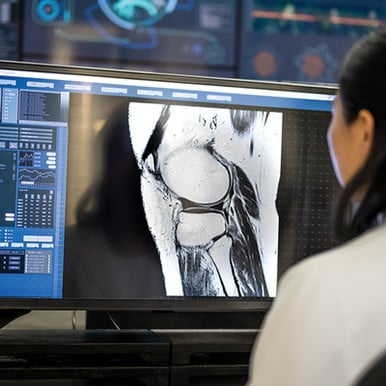
- Radiography of Knee Injury – Correlation with CT and MR – Mini Pathria, MD
- MRI of the Knee – Meniscus and Cruciate Ligaments – Kevin Sweetwood, MD
- Arthritis – Beyond Black and White – Kevin Sweetwood, MD
- Metabolic Disorders – Daria Motamedi, MD
- Extensor Mechanism of the Knee – Daria Motamedi, MD
- ACL Graft Reconstruction and its Complications – Daria Motamedi, MD
- Multimodality Evaluation of Tendons of Wrist and Hand – Kevin Sweetwood, MD
- MRI of Shoulder Trauma – Kevin Sweetwood, MD
- Pitfalls in Shoulder MRI – Daria Motamedi, MD
- Hip MRI – Labrum, FAI, and Beyond – Daria Motamedi, MD
- Problem Solved – Applications of MSK US – Daria Motamedi, MD
- MR of the Pelvic Tendons – Mini Pathria, MD
- MR of Muscle Injury – Mini Pathria, MD
- MRI and US Correlation Cases – Kevin Sweetwood, MD
- Ankle and Foot – Commonly Overlooked Injuries – Mini Pathria, MD
- Stress Injury of Bone – Mini Pathria, MD
- Skull Base Lesions – Christine M. Glastonbury, MBBS
- Headache – Do Not Miss Diagnoses – Yi Li, MD
- Top 3 DDX in Head and Neck – Christine M. Glastonbury, MBBS
- Work Up of Non-traumatic Intercranial Hemorrhage – Yi Li, MD
- Imaging the Neonatal Brain – Yi Li, MD
- CSF Pressure Disorders – What the Diagnostic Radiologist Needs to Know – Vinil N. Shah, MD
- Emerging Concepts in Hydrocephalus – Yi Li, MD
- Imaging of Pituitary and Hypothalamic Abnormalities – Yi Li, MD
- Spine Reporting Essentials – How to Add Value – Vinil N. Shah, MD
- Autoimmune Encephalitis – Yi Li, MD
- Spinal Cord Inflammatory Lesions – Vinil N. Shah, MD
- Post Surgical Changes in Head and Neck – Christine M. Glastonbury, MBBS
- Spine Tumor or Mimic – How to Tell Them Apart – Vinil N. Shah, MD
- Salivary Gland Lesions – A Case-Based Review – Christine M. Glastonbury, MBBS
- Spinal Fusion – Postoperative Spine Simplified – Vinil N. Shah, MD
- Pearls and Pitfalls of Head and Neck Tumor Imaging – Christine M. Glastonbury, MBBS
- Challenging Spine Cases – Lessons Learned – Vinil N. Shah, MD
- Errors Are Opportunities – Thinking About QA a Different Way – Christine M. Glastonbury, MBBS